Field of Science

Showing posts with label microscopy. Show all posts

Quick ImageJ Tutorial: Scalebar calibration

Due to popular demand, I'll sporadically write up a series of posts on some ImageJ basics. For those not in the loop, ImageJ is a popular open source image processing program for microscopy; due to the availability of some rather sophisticated plugins, it can be a very powerful tool in the right hands. Also, the customiseable shortcuts are a blessing (and a curse for one's muscoloskeletal system, especially in the wrist region). I'm by no means anywhere near an expert with ImageJ (and don't do anything sophisticated, like macros or even much 3D/4D work), but there's a few basic things that I had to learn, and would like to share with anyone interested.

I put the rest of the post under the fold as most of you probably don't care...

Pond Microforay part 2.5

Preceded by parts 0.5 and 1.5. This time in decent DIC!

Another Cyclidium with a sail-like 'membranelle' - the haplokineties of the oral ciliature. I kind of like this picture...
The story behind 'membranelle' is that this row of cilia behaves much like a membrane would, thus leading microscopists of the past to consider is as an actual membrane. Later it turned out the 'membranelle' has nothing to do with membranes, but the name kinda stuck. I've had an instructor who was viciously against 'membranelle', and despised the use of that word. Same instructor also hates 'protozoa', so I learned to be very careful with that word...


A collage of random flagellates (and a spore-like thing). Feel free to identify A,B and D.

A random amoeba. Discosea?

Coleps, the "hyena of the protist world"! They will eat pretty much anything...

Cyst, perhaps of Actinophrys sol ('heliozoan'). Which can also be apparently violent enough to engulf an entire Colpodium(ciliate).

Collection of more heliozoans, likely centrohelids.

I kind of like the bottom left one...

Almost definitely a centrohelid, which are now with a Crhaptophytes/Kraptophytes*. Note the messy spicules near the cell body. Also note the centroplast in the middle. The axopodia (long thin threads sticking out) are microtubule-based structures which are used for capturing stuff (via kinetocysts - the little blobs on the threads); they can also contract rapidly (1/30 s!). This may well be Raphidiophrys, but I can't say for sure.

*I now have the story behind this. Apparently, they wanted to name the group 'Craptophytes' in one of the papers, but a reviewer rejected it because it didn't include the Katablepharids in the name. They considered changing it to 'Kraptophytes', but got worried that the name might actually go through, and may even set a rather questionable trend... so they named them 'Hacrobia'. Colloquially, however, people still often refer to them as Cr(h)aptophytes. There was an 'h' in the version told to me by a postdoc, so I now have a choice between the following: 'Craptophytes', 'Kraptophytes' and 'Crhaptophytes'. The latter looks the weirdest, and blatantly includes the Haptophytes. And the 'crap' is a little more subtle. So I'll keep using Crhapto.

And finally, a random metazoan. Daphnia?

And that's it for this particular microforay. Got loads of own microscopy to do tonight, maybe I can sneak in some more pond samples too while I'm at it ^_^

Pond Microforay part 1.5

More pond pictures (part 0.5 is here). The first hour or so was dedicated to putting up with very shitty DIC because some moron decided to 'frobnicate' with the bottom polariser, which is not meant to be touched very often. Luckily, I eventually managed to out-'twiddle' their frobnication and adjust the scope properly. In that first hour I did find some cool stuff, so please excuse the embarassingly shitty image quality. The polariser was misaligned, grrr!

So here's some Difflugia-like amoebozoan. They're amoebae which ingest and secrete random particles to make a test. (sometimes, those can even be diatom valves!)

A random amoeba. Don't expect me to ID amoebae... especially with shitty lightning! (still sore about that, yes!)

Then the slide was drying out, so I decided to add a drop of water.

Everything stopped moving, and finally I got a shot of a euglenid (remember that our camera is painfully sloooow!) By stopped moving, I mean I could even get an awesome shot of a flagellum, and a badly deformed cell (another euglenid):

By now I was confused. I look at the water dropper - it's labelled 50% glycerol. Shit. Who knows what was actually in it, as it induced a diatom to blow up rather impressively:

Somehow I doubt glycerol is supposed to do that. Probably screwed up the osmotic pressure drastically - I'm guessing typically one uses ~10% glycerol to slow things down in microscopy? Anyone know? Plants don't need slowing, so the glycerol there is added to help match the refractive index more than anything else...

Made a fresh slide. First off, Difflugia-like amoeba in mitosis(? or not...do they redistribute test material during division?):

(REALLY sore about the shitty image quality - this would've been so amazing under proper DIC!!!)
Something vaguely looking like an Actinophryid... (although not as vacuolated as it should be, but who knows...)

And what appears to be an abandoned Bicosoecid lorica:

And then I had an encounter with what appears to be a dividing ciliate!

Damn I want my DIC by this point. A ciliate in mitosis - how cool is that!? Grrrr! You could even see the oral ciliature! Can't wait to work on ciliate morphogenesis/development (if they're crazy enough to let me in, that is...)

Speaking of ciliates, the armoured Coleps:

A few random amoeboid things:

Random flagellates: (there were a lot of Cyclidium (ciliate)):

And I really need to brush up on my euglenids!

Being beyond fed up with the lack of DIC, and knowing our scope is actually quite good, I finally twiddled my way to the problematic polariser. Suddenly, life was good again!

"HOLY SHIT, IT ACTUALLY WORKS NAO!" I even allowed some blatant overexposure to creep in... >_>

So here my images actually started to suck less. And more cool stuff was seen. Now that we're all waiting in suspense for marginally acceptable microscopy, I'll call it a day here. Stay tuned for part 2.5: This time with DIC! ^_^

Termite gut microforay - excavate time!

A couple days ago a local basal winged termite crossed my path. Then, like two microtubules approaching each other below the threshold angle, our paths zippered up together and the poor sucker had its parabasalian jewels strewn exposed on the slide. No worries, the termite lives on, albeit as a carbon source for other life...

Now I have rather limited experience working with gut endosymbionts (my second time), so I suck. Also, I didn't have the right solutions for keeping the denizens from exploding osmotically. So my images suck too. Kind of like this poor exploding trichonympha:

Actually, they seemed to be less exploded later when mounted in 5% glycerol (or perhaps they managed to die in the termite while I was working on the first sample, and experienced less osmotic shock that way somehow...) The glycerol may have helped out in the refractive index matching a bit, although perhaps I just magically came across better DIC settings later... (I need to stop speaking in strings of hypotheses. [fundie]I believe the glycerol helped my imaging, and I shall guard my faith until forever! Yeah, take that, 'evidence'! In other news, termite endosymbiosis is older than the earth itself. [/fundie])

First thing I saw was some mystery nematode: (was wiggling about way too much for a decent image) How do you key out nematodes anyway???

Top right: Some mystery...thing. Looks like an ex-organism, judging by the characteristic bubbles that tend to form when anaerobic protists die. Then again, those may well be artefacts of something else. Any ideas?
Bottom:Trichomitopsis! (it actually looks really cute when not completely mutilated)

(the scalebar on the second one is wrong; that would be about 12.5um, forgot to adjust for 40x settings...)
A slightly better shitty image of streblomastix; how many flagella can you count at the anterior end?

And fresh from the Crap on the Bottom of the Slide department, some mystery flagellates(?), or pieces of dirt sitting on top of bacteria (Or elongated pieces of crap). Hey, identifying whether crap is/was alive or not is kind of difficult sometimes! Although I'm pretty sure (B) is a real thing. (D) also looks kind of flagellated... any ideas?

Edit 18.08.09: With Opisthokont's help, possible IDs: A - hexamita?; B, E - monocercomonoides; C- a thing with an axostyle, or hexamita)

Then I had fun with the carnival ride that is a prism and and couple polarisers (aka DIC):

Trees in the autumn sky...

...roots on the autumn ground?

Shiny!

Omg, pseudo-darkfield! (no it's not)


The helical spiraly thing is a piece of xylem, methinks; termites eat wood right? Well, trichonympha eat wood inside the termite... so you get pieces of plant matter in them. And then you find random stomata floating about, which I'm pretty sure are not some weird contamination from my arabidopsis screening stuff (kinda reused the slide...)

Those things freaking haunt me, even when not doing research! Amazing how it survived intact...stomata can be quite fragile. Although perhaps that's specifically when you need them not to be...
Actually, wait, question: how the hell do stomata get in there if termites eat wood? While plants do have stomata on their stems, 1. termites don't munch on epidermal tissue all that muchl; 2. I doubt woody plants have any once bark is formed. Maybe it -is- some weird contamination from the previous sample...luckily, not actually doing any real science here.

Speaking of stomata, I has works to do. We're kind of like plant dentists - working on 'mouthes' all day! Especially with gene names like four lips(FLP), too many mouthes(TMM), speechless(SPCH), mute(MUTE), moustaches(MUS), etc. We also have YODA. Apparently somewhere in the Arabidopsis genome lies a regulatory gene SUPERMAN and its suppressor KRYPTONITE. We like to keep ourselves entertained...[/derail]

Let me know if you want more 'microforays'!

Leaves on drugs

I accidentally hit Shift+I in ImageJ, which inverts colours. The result was very trippy:

(originally an Arabidopsis leaf surface; yes, drugs have been involved - for once, on plants rather than from plants...)
Seriously, doesn't this almost look like some modern abstract painting thing?

Going back to working on the carpal tunnel syndrome quantifying microscopy data...

Btw, I proudly use ImageJ for most of the cropping/editing work in this blog. I am not a nerd. To prove the latter point, I should perhaps someday have a post dedicated to revealing the inexhaustible Awesome hidden in the ImageJ universe. We microscopists have a freaking fetish for it... >_>

Naughty nomenclature + Deconvolve This!

Taxonomy can actually be fun sometimes:

Tubifex longipenis

heehee... somehow there's this public image of academics being all mature and serious. In reality, those are rather hard to find...


Recent disappearance was due to leeching off a 2wk 3D live cell microscopy course that happens at UBC every year; they were nice enough to let local students attend lectures... and even leech off the food a bit... Great course, highly recommended for those who think microscopy is just putting shit on a slide and looking at it. Microscopy is a high art and a rather sophisticated science. Course is likewise quite recommended for those who realise that already...they have pretty toys cutting edge microscopes to play with test out for your research purposes.

So yeah, practice safe microscopy -- always use deconvolution on 3D data*! ^.^

Also, physics (and even chemistry) becomes surprisingly interesting when presented by the right people in the right context! Or maybe I'm just crazy... (but so are the course faculty and many students so no one noticed...)

*There is a debate on whether deconvolution works at all on plant material, which is rudely autofluorescent and absurdly packed with spherical aberrations sufficient to utterly destroy your PSF into a disturbing calculation-intensive mess. Our stuff is apparently notoriously shitty for decent microscopy, especially in confocal and multi-photon. And guard cells are among the worst in plants... so decon may not work there (Sorry, Prof. Pawley! >_> )

Science as a journey through alien worlds

Running a gel, thus perfect time to blog. Perhaps if I write stuff in the lab, it may actually turn out to be marginally intellectual.

Hopefully my gel won't turn out to be something like this again:


I hate genotyping - the work itself requires the intelligence of a brain-dead foetal monkey, yet manages to find a stunning variety of ways to fail. And when it does work, it usually shows everything was wildtype anyway. Or maybe your mutant-specific primers failed. You should probably remember to run a positive control next time. If you have one, that is. And when it does work, you can brag about find a homozygous line amongs the seeds you ordered from the stock centre. Nature-worthy achievements right there.

Actually, most lab work is about as mundane and intellectually uninvolving. The planning and literature reading, data analysis and troubleshooting(!) can be quite exciting and tend to involve plenty of thinking, but you only do that in-between the endless repetitive pipetting and mixing chemicals and following protocols. I think research probably involves one of the biggest intellectual (and emotional, heh) rollercoasters of all professions, which is why only the very insane can handle it.

[did I run the DNA off the gel yet... no, phew.]

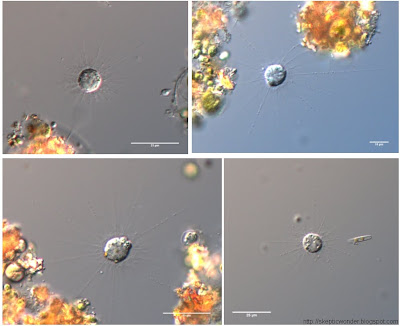
I do enjoy microscopy, however, which is considered by some to be repetitive and mindless as well. I cannot disagree more. It's an artistic break from the gels and protocols - you have a lot of factors to play around with to obtain the best image you can. Microscopy is also very dangerous, a wonderful way to obtain false positives! It's extremely easy to tweak the image to see what isn't there, even unintentionally. A substantial component of the art is to obtain sexy images without sacrificing the scientific validity thereof.


As children, many of us have dreamt of fantasy alien worlds, to be much disappointed upon finding out we would most likely never encounter any; not within our lifetimes anyway. However, it is wonderful to know that alien worlds are not confined to what lies beyond the Earth's atmosphere! (or present time...) They are all around us, ready to provide enchantment and fantasy to anyone who invests enough to explore.

To a mathematician, beauty may lie in quantifying forms or dynamic phenomena, or working with abstract worlds far beyond the imaginative capacity of the human mind. To a fiction writer, fantasy worlds thrive in his mind not much different from the form taken by reality itself. To an astronomer, the alien worlds are almost within reach - just barely visible, yet not ours to manipulate. For me, foreign worlds exist under the lens of a microscope, where if you magnify anything far enough, you can spend a lifetime wandering amidst the surreal landscapes unseen by naked eye. The life of or within a cell is fascinating enough conceptually, but that mysterious aspect of it being beyond our 'scale' makes it not much different from a childhood fairytale or a sci-fi novel - except that it's real!

On a more abstract level, science itself is a foreign world for exploration (along with math, the arts, etc.). Upon liberation from what Carl Sagan calls "the anaesthetic of familiarity", the mundane everyday world around us becomes full of tantilising mystery. Science is a way of unraveling some of this mystery (I'll argue, the optimal way of dealing with natural mystery), an ardurous journey through the unknown worlds. It's rather fractal-like in nature - the more you explore, the more questions arise; the deeper something is examined, the more detail beckons attention.

I think a good analogy for the scientific journey could be hiking or climbing - the act of walking along a trail is quite repetitive: all you do is place one foot before the other. After some time the scenery begins to repeat itself, and one wonders how anyone could enjoy outdoor activities at all. But many do, and probably few ever notice the monotony of walking, or climbing, or paddling a kayak... for there's always something fresh to look at, something wonderful to appreciate and enjoy. It's fairly easy to knock off the 'anaesthetic of familiarity' for an outdoor enthusiast - the escape from the chaos of the human 'world' is sufficient to make the journey worthy of the physical investments. The threat of an occasional failure, the pain of stepping on a too-unstable rock... is usually not enough to deter one from actually enjoying the exploration itself.

Science is quite similar - there is the monotony and rigorous exactness required by the scientific method, the endless protocols, the multitudes of failed and inconclusive experiments. There is the threat of losing one's job upon failure of publication - the world of 'publish or perish'. It seems one must be insane to voluntarily engage in such activity, much less enjoy it. But just like the monotony of walking is instantly forgotten upon the sight of a rare bird or an overpowering landscape, the monotony of science is instantly lifted upon the sight of a contradicted null hypothesis, or the sight of a rare species, or the ability to actually see a cellular process in action in real time... (perhaps for a chemist, the synthesis of a brand new chemical never before produced on earth could induce a similar rush or euphoria?)

But just as one does not go on a hike expecting to see a rare bird (lest the foiled expectations lead to a wrecked mood), one cannot go into science expecting to make great discoveries. The very process itself must be tolerable, and hopefully captivating enough, to keep going. Sometimes you get excited about the potential outcomes of an experiment, only to discover your results are utterly inconclusive and the story is far more complex than previously imagined. I think what makes science quite suitable for an insane type of mind is the ability to actually not mind such disappointments, and savour them as a testament to nature's superiority and infinite complexity.

After all, we may rest assured that we will never run out of work to do, just as an outdoorsman would never run out of places to explore. Now isn't that a comforting form of eternity?


[genotype results: all wildtype. Time for more PCR!]

Delicious squashed cockroach guts

Done finals for the term. Have more in June, so it doesn't seem like the end really...

And a certain Dictyopteran-endosymbiont-obsessed faculty in our department is indirectly responsible for the following 'study break':

I'm sitting there, cramming biochem, getting some studying done. Along comes a cockroach (I've spotted at least three species of cockroaches in our building already). I instinctively catch it and key it out, to subsequently determine it's phylogenetic position without the cockroaches. It was Supella longipalpa, sadly not a basal roach, and thus unlikely to host the Parabasalian zoo I was looking for (eg. Trichonympha). Further literature searches revealed only bacterial gut flora, and parasitoid wasp larvae. However, anything trumps biochem, so I decided to go have a look.

As expected, not much excitement in the way of gut flora. A couple flagellated-seeming tiny things (a couple microns) -- possibly bacteria, possibly some obscure excavate -- couldn't quite tell. Couldn't take pictures of those as our camera is too slow even for us plant people. But then I discovered the awesome that is insect tissue under polarised light:


Microscopy is truly the closest thing we got to exploring foreign universes...


Surreal landscapes...


This one reminds me of Southern Ontario fields in the winter:

(you can see why cockroaches are in order Dictyoptera - net-wing.

More Martian tripiness:


And can anybody tell me what those greenish round things are?


I really need to read up on insect anatomy... have no idea what I'm looking at most of the time. Not to mention that it's all squashed to begin with...

Sadly, as expected, no cool Parabasalian freaks found in Supella longipalpa hindgut this time. K, next cockroach!

If anyone's got some spare Cryptocercus lying about, feel free to send 'er over! My current clip of Saccinobaculus sucks arse, and that organism is way too cool for sucky videos.

Plant skins enlarged

I was down with a cold lately, along with a couple midterms, so appologies for crappy posting. Since I don't have time or energy to write anything thoughtful at the moment, I'll shower ye with some random pics from my collection:


Liverwort leaf surface -- one cell layer thick, round cells


Bladderwort (aquatic plant) with permanently closed stomata -- relics of the plant's terrestrial ancestors.


Arabidopsis leaf; UV fluorescence: DAPI (binds to DNA and stains blue) and chlorophyll (red) autofluorescence. Those two huge things are... drugged up mutant stomata! The tissue was overstained and too old, thereby sucking for science, but it made some pretty nice desktop wallpaper material!

And just because I love how UV imaging makes everything seem surreal :


It's almost a bit nebulous, isn't it?

Not digitally altered in any way -- looked just like that in the eyepiece!


Sigh. I am married to microscopy... can't help it!

And then it suddenly... EXPLODED!

An image sequence of a cilliate I got today, while taking optical sections:


(40x, Namarski phase contrast)

The heat from the light source must've got to the poor guy... =(