Field of Science

Showing posts with label amoebozoa. Show all posts

Dermamoeba – Having your coat and eating it too

This post was chosen as an Editor's Selection for ResearchBlogging.orgWe've been neglecting the micro-squishies lately (filose amoebae ain't proper squishies – too many fine protrusions in the way). Amoebozoa is a eukaryotic supergroup comprised of predominantly lobose amoebae, meaning their pseudopods are rounded and not fine and pointy (like those in the preceding post's organism – Filoreta). Aside from the test-bearing Arcellinids, amoebozoans tend to be naked amoebae ('gymnamoebae'), like the well-known Amoeba proteus, often erroneously referred to as a 'primitive', 'simple' or 'ancient' organism. "Naked amoeba" is a bit of a misnomer – while they don't lug rocks and heavy dishware around like testate amoebae, they generally carry some sort of cover, as most cells do. Gymnamoebae just pack light. Some, like Cochliopodium, dress themselves in intricate scales, while others, like many Vannellids, are covered in thin, pointy glycostyles. Dermamoeba, in turn, wears a thick, heavy coat.

5-8 Dermamoeba going about its business (n – nucleus, cv – contractile vacuole). 9 – Dermamoeba lounging about in cysts (c) upon devouring some algae (chain-forming diatom or some Trebonema-like thing). Nom nom nom. (Smirnov et al. 2011 EJP)

Dermamoeba's fine coat consists of thick bi-layered glycocalyx (a covering of fluffy sugar-proteins), sometimes with additional 'dense matter' lining the cell membrane. Upon encystation, an extra layer, the cell wall, is formed, but the contraption is thick enough without it already, at about half a micron.

EM sections through the intense Dermamoeba cell coat. m – cell membrane, gl – glycocalyx, adm – 'arrangement of dense material' (ie, "we don't know"). The glycocalyx often forms pretty patterns when sectioned. (15 is part of a Golgi body) (Smirnov et al. 2011 EJP)

This thick coat poses some problems of its own. Amoebae eat by engulfing prey with their pseudopods – and this involves some degree of nudity and cell membrane exposure. Half a micron of glycocalyx wouldn't be particularly flexible, and and not much fun to digest. Dermamoeba has to nibble on its coat before the meal. Upon contacting prey (typically algae), the amoeba forms a concave food cup around it, from the centre of which the cell coat gradually disappears. As the food cup deepens, the prey is pulled in to meet its doom via thick bundles of actin microfilaments spanning much of the cell – another unusual feature of this process. The prey is consequently engulfed for eventual digestion. As a result, the prey-containing vacuole has no glycocalyx for the amoeba to choke on (or rather, presumably, waste energy digesting).

Diagram of Dermamoeba's unusual feeding procedure. After the algal prey (al) is contacted by the amoeba (am), the glycocalyx (gl) is digested and the prey is drawn in by thick actin microfilament bundles (mf). The resulting food vacuole (fv) is conveniently devoid of coat material. (Smirnov et al. 2011 EJP)

And here the food cup is 'live', or was before some electron microscopist brutally murdered it in osmic acid and sliced it up:

EM sections through prey (al) being engulfed by the amoeba (am). Note the disappearance of the glycocalyx (gl) at the centre of the invagination. (Smirnov et al. 2011 EJP)

How do some of the other coat-bearing amoebae get around their irremovable clothing? Without going into much detail (amoebozoan surface coverings are really cool...), the glycostyle-bearing Pellita simple sticks small 'subpseudopodia' through it for both moving about and feeding. In fact, some propose that the glycostyles may help it move by reducing the surface area in contact with the substrate – keeping the sticky cell membrane away on stilts.

Top left: Pellita walking on stilts of glycostyles (depicted at the right). Bottom: extruding sub-feet across stilts for feeding. (Smirnov & Kudryavtsev 2005 EJP)

I'm decidedly avoiding amoebozoan systematics here. Christopher Taylor did a nice overview of it at the Catalogue of Organisms a while back, but keep in mind that some of the groups did jump around since then, and the phylogenies are in the works. Maybe if more people cared, the taxonomy could be resolved sooner...

PS: My committee* has voted to remove "Sunday Protist" from Sunday Protist titles, since:
a) They seldom come out on Sundays anyway (lulz); and
b) Takes up too much valuable headline real estate. Since we bloggers are supposedly playing pseudo-journalists or something, might as well play it right... ;-)
(and c) Structure and I aren't the best of friends.)

* Given how inefficient my brain is at accomplishing anything, I've concluded it can only be composed of a close neural approximation of a committee. Explains the indecisiveness as well. Probably requires a double majority to pass any major decisions, and hence is about as effective as the Californian government. Without the sovereign debt crisis, fortunately.


References
SMIRNOV, A., & KUDRYAVTSEV, A. (2005). Pellitidae n. fam. (Lobosea, Gymnamoebia) – a new family, accommodating two amoebae with an unusual cell coat and an original mode of locomotion, n.g., n.sp. and comb. nov European Journal of Protistology, 41 (4), 257-267 DOI: 10.1016/j.ejop.2005.05.002

Smirnov AV, Bedjagina OM, & Goodkov AV (2011). Dermamoeba algensis n. sp. (Amoebozoa, Dermamoebidae) – An algivorous lobose amoeba with complex cell coat and unusual feeding mode European Journal of Protistology : 10.1016/j.ejop.2010.12.002

Reticulose amoeba: cells can be fine nets too

Again, the protist kingdom is a special paradise for a cell biologist: as soon as one steps outside the plant and animal kingdoms (and yeast), diversity of cellular forms and structures explodes beyond reason. Cells can also take the shape of a fine net with no obvious cell body proper:

Cover slip floated ~ 1wk on marine sample from intertidal silt at Stanley Park. (40x obj, DIC and phase, resp.)
EDIT: Confirmed Filoreta.

Almost overlooked it thinking it was just slide gunk. Amoebae suffer all too often from that fate – apparently Parvamoeba, one of the most common and ubiquitous amoebae, was only described in the early 90's (Rogerson 1993 EJP) because it was tiny and no one noticed.

Could be something like Filoreta sp. (Rhizarian), but something feels off about it. Filoreta doesn't seem to stretch cytoplasm between filopodia like this specimen does. Maybe it's more like the amoebozoan Corallomyxa and Stereomyxa, or stramenopile Leukarachnion. Then again, amoebae are notoriously dynamic in their morphology. Something that's a far bigger issue in the microbial world is the necessity of getting a sense of the morphotype range of a species; one specimen doesn't quite cut it as it does for animal taxonomy.

In fact, perhaps instead of the ridiculious (for us) ICZN and ICBN requirements for submission of material for curation (many species neither like being cultured nor preserve all that well on a slide), for microbial species there should be a requirement for additional images of different specimens, if possible, to try to capture some of the morphological range. But then again, I'm not a taxonomist, so what do I know.

Right, midterm... (hey, at least I procrastinate productively!)

Gluttonous amoeba

Was looking through some freshwater pond samples one day, and saw this amoeba (Polychaos?) which has managed to engulf an entire Phacus (a photosynthetic euglenid)!

How it managed to ensnare the Phacus with its pseudopodia is beyond me. Phacus is perfectly capable of swimming away. Then again, amoebae are actually kinda scary...

Or what if this euglenid wasn't actually being digested, and would have eventually become a permanent endosymbiont, forming a brand new lineage of photosynthetic amoebozoans with tertiary plastids? And just as this miracle was about to happen, some asshole biologist captures the poor amoeba and kills it. Oh well.

Moss Microforay Part 1: Assorted critters

Posting and attempting to identify own micrographs is a quick and easy way to fill up blog posts when one is short on time. Aside from all the time spent at the scope and processing the images (ImageJ, baby!) of course. At least there's no expiry date on these, unlike hot new topics which need to be blogged within an hour else the entire blogosphere will scoop you, somehow...

Moss was sampled on a coastal mountain on the San Francisco peninsula. As the sampler is a labrat and therefore not particularly sample-savvy, said specimen was abandoned for three weeks only to be rehydrated a couple days prior to imaging. Thus, there is a high probability that much of the really cool stuff died off before I got around to it. Conversely, some of that cool stuff may be damn good at encysting, and is now perhaps freshly-excysted.

Saving the Euglyphids for the next installment. Mwahaha. Let's go over an assortment of random non-Euglyphid organisms first. It would be nice to get some help with ID from the experts lurking here *wink*

Ok, first off -- flagellates. That is, glimpses of the few flagellates that weren't moving so ridiculously fast they were but a blur on the picture. There seemed to be a lot of bicoesid-like loricate things (or vaguely look like them anyway):

That cell may have been damaged somehow, or is it actually being amoeboid-ish? Can bicoesids do that? My understanding is they tend to be rather rigid, but I'm not aware of any other loricate flagellate besides Dinobryon and choanoflagellates. This thing is definitely neither of those.

This specimen appeared attached along the length of the flagellum at the top of the picture.


This one looks more like a bicoesid. As do the next three:


This one too, probably, though the lorica can't be seen. There are aloricate bicoesids as well.

Bicoesids are actually borderline suitable for microphotography with a slow camera due to their habit of attaching to things. Seriously, sessile organisms are really nice that way...

More potentially cool flagellates that were moving around too much to get a decent shot:


This was a sequence following a strange amoeboflagellate-looking thing(s) -- any idea what this may be? A cercomonad of sorts?

Same cell(s) in all of the images. Was shot in a short timeframe (maybe a minute or two), so highly doubtful that cell divison could've occured within that time. Or maybe not?

Amoebozoan time! May ever try ID'ing some...before doing that, I feel compelled to draw your attention to Smirnov & Goodkov 1999 Protistology, a wonderful overview of and reference for amoeboid morphotype diversity -- amoebae do have shape! Another good source for ID'ing amoebae is Smirnov & Brown 2004 Protistology, and an associated site (though sadly not maintained since 2003ish =( ); I will be relying on those to see how far I can go:

This one seems to be flabellate with adhesive uroidal filaments; may be Flabellula (something off about that), Stachyamoeba (perhaps? esp. this picture); Rosculus seems to lack prominent uroidal filaments as well as seemingly a Heterolobosean from what I've gathered. Could also be of the fan-shaped morphotype but those don't seem to leave trailing adhesive filaments that much. I'll go with Stachyamoeba for now, hopefully no one finds that offensive...

Ok, the next one is probably polytactic or dactylopodial by morphotype.

Candidates: Amoeba (too small), Chaos (doesn't seem multinucleate and huge), Deuteramoeba, Thecochaos (not wrinkly enough), Polychaos (something meh about that too). As for dactylopodial candidates, Paramoeba is looking better, as is Korotnevella. As the former is much more commonly mentioned, I'll go with Paramoeba for this one.

By the way, the prominent round thing in the middle is most likely the contractile vacuole; note how it obvious changes size during optical sectioning, especially in the first specimen. Also, amoebae aren't always amoeboid -- many species take upon 'floating forms' which tend to look like spiky balls. Some amoebae are apparently quite at ease with being planktonic too, like this vicious rotifer-munching Difflugia mentioned earlier.

Amoebae can actually move pretty quickly -- this one is fuzzy due to motion blur.

Morphotype: Lens-like (Smirnov & Brown 2004); candidate: Cochliopodium (and here). Really does look like Cochliopodium; perhaps I got at least one right!

A few more fun amoebae:

In the last one, note how the clear pseudopodia emphasising the contrast between ecto- and endoplasm -- the latter being fairly gelatinous and containing the 'stuff' of the cell; the former, full of actin, responsible for cell motility.

Arcellid amoeba: (proteinaceous test -- note texture)


Odds and ends: First off, ???:


What I think might be a developing nematode egg case (elsewhere there were very similar things with nematodes inside):


Some fungal thing? (germinating spore?)


And last but not least for today, a bacterial filament:


Ok, back to cramming...stay away, internet! Must know everything there is to know about invert biol by Monday...aaaah~! Also, apparently metazoan phylogeny is in about as much of a mess as the the Tree of Eukaryotes. Kind of creepy...

Fossil testate amoebae

There's something about the idea of fossilised single-celled organisms that's just pure awesome. Even if it's just their shells.

For example, take a look at these past relatives of Centropyxis and Leptochlamys from Schmidt et al. 2010 JEM, AOP:

Testate amoebae from 100mya amber in France. The arrow in 1 points to what the authors believe may be fossilised cytoplasm flowing out of the cell. 2) four ventral pores visible. 4-7) holotype of modern Leptochlamys, optical sections. 8-11 potential resting cysts. All scalebars = 20um (Schmidt et al. 2010 JEM)

Curiously, unlike the representatives of modern genera, these amoebae have perforations in their shells. Now, the very resemblence to Centropyxis may well be a case of convergence, as it's not that unusual for an amoeba to evolve a test one way or another - Euglyphids, for example have nothing to do with amoebozoa, and yet have rather elaborate tests as well. But provided these specimens do originate from the same lineage as modern Centropyxis, and provided these perforations are real, and not just holes caused by some predation or fossilisation artefact (their asymmetrical arrangement raises some questions...), it shouldn't be surprising that amoebae have not been in perfect stasis for the last 100 million years (or even more, as is commonly assumed).

On one hand, amoebae are pretty good at what they do, and thus could probably continue surviving well as they are for another few hundred million years. Their large population size should buffer them from excessive drift, and perhaps there isn't as many possible viable ways of being in the 'design space' for an amoeba, though the latter assumption is extremely dangerous and probably very wrong as there seems to be no limit in all the ways those seemingly 'simple' organisms can utterly stun us. But there may be something to it, just combinatorically speaking -- there are more possibilities if you're big and full of junk, like metazoa.

On the other hand, things like amoebae may have had as violent of an evolutionary past as the more famous multicellular creatures. We're not particularly sensitive to variations in structures beyond our familiar scale, especially considering those tend not to fossilise well. So it may well be that a) many of the fossilised modern-looking testate amoebae are actually completely unrelated (if we have issues with morphological classification leading to polyphyly even in modern taxa...!) and b) there have been unimaginable diversity spawned and respawned and extinguished in the past that we simply cannot detect due to the rather crude methods available to us.

What I'm trying to point out is that it is an error to automatically assume that "lower organisms" (*twitch*) are in some sort of long-term evolutionary stasis, as is so commonly done. Of course there are things in stasis, and of course some organisms have had more violent evolutionary histories than others (eg. parasites, especially intracellular ones), but it is probably unwise to predict that based on the 'simplicity' or size of an organism, or, worse yet, how distantly related it is to us. Unless good data supports that, of course --please let me know if such data has been looked at!

Sigh, just as it looks like those of us working with extant organisms have it pretty bad, figuring out even the basic questions becomes so much harder for the paleontologists who only have questionably preserved fragments of the past to look at. It's truly amazing we can even begin to reconstruct any of the past at all! And that is why it drives me furious to hear comments like "We weren't there, so we'll never know what happened in the past, so why should we care?" Personally, I can't figure out what the hell is going on in the present either...

Reference
SCHMIDT, A., GIRARD, V., PERRICHOT, V., & SCHÖNBORN, W. (2010). Testate Amoebae from a Cretaceous Forest Floor Microbiocoenosis of France Journal of Eukaryotic Microbiology DOI: 10.1111/j.1550-7408.2010.00471.x

Of random rotifers and vicious amoebae

ResearchBlogging.orgStill procrastinating with the Heterolobosea posts (no, I haven't forgotten). Real protistologists give me sorry looks when I mention being stuck writing about that rather obscure and messy group. I don't want to just do a taxonomic overview - I like to mention odds and ends about their cell biology as well. Sadly, the cell biology of most of those things is a sorry neglected mess. Even the eruptive pseudopodia that are quite characteristic of this group (although present in some amoebozoans as well) seem to not have been examined on a molecular level - I cannot find a single paper describing the cytoskeletal dynamics behind this peculiar mode of motility, and my sources point out a severe shortage of attention in that area. But I'm still scraping things together, so eventually something will appear...

Now for something completely different...since someone just sent me scouring piles of archaic metazoan 'mesozoan' literature to identify 
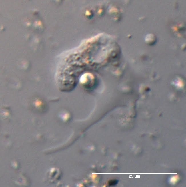
that damn bug (an orthonectid of some sort), I have a random metazoan of my own to show off a bit, from the latest pond microforay:


Rotifer. (Mine =P 40x* DIC) Note the contractile vacuole-like structure (actually its bladder, but appears to function in a very similar way. Another case of ultimate convergence?).


I can't quite key this one out, especially since this damn sucker is on its side. Probably the only reason I was able to capture a photo of it, since they tend to be quite hyperactive normally. This one appears to be immobilised by the cover glass. Perhaps it may be Aspelta or something from the Lepadellidae. Or might it be a Bdelloid? Perhaps a certain taxonomist could help out. Especially the one who made me browse orthonectids for the past few hours. ^^

*40x here, and onwards, refers to objective rather than magnification. We don't actually care about mag, it's the other properties of the optical system that matter much more when dealing with professional instruments. (Such as numerical aperture and field of view, focal length, focal depth, which type of immersion medium its calibrated for, etc)
Technically, I should be writing down the NA and such, but we're not that meticulous crazy around here.
**Randomly found out there's a
rotifer that lives inside colonies of Volvox, as a parasite...


Much of my resentment towards metazoa simply comes from them being overstudied and overemphasised, to the neglect of other phyla. That said, they are still interesting and still poorly understood lifeforms, especially among the spineless things. While representing but a mere sliver of life's overall diversity -- contrary to what our senses tell us -- metazoa are nevertheless, like all life, fascinating and weird. Metazoan multicellularity, as mundane and ubiquitous as it may seem to our rather biased eyes, is about as weird and insane as kinetoplastid kDNA editing, or the convoluted ciliate nuclear genomes, or the breathtakingly massive, and seemingly pointlessly so, nuclear genomes of dinoflagellates and some amoebozoa. I'm quite disappointed at the absense of the 'perspective of strangeness', if you will, that seems to dominate many animal-oriented classes (especially those catering to pre-meds. Must. not. rant.)

Interestingly, off all taxonomic and phylogenetic literature, I find metazoan papers to be the most difficult to follow* - we actually have common names to refer to quite a few animals metazoa. Furthermore, the obsession with ranked hierarchy is driving me insane - you DO NOT have to name every single possible grouping you can come up with, seriously! That makes searching for stuff an absolute disaster, as you have to look for the relevant subfamily, family, superfamily, suborder, order, superorder, hypersuperorder, subclass... etc. All named differently enough to not fit into one convenient Google (or PubMed, or WebofScience) search. And you have to find all of them. And they change. And your life becomes a living hell. Please stop, seriously! WHYYYYY!? Did TC-S write their taxonomies or something?! And they listened to ALL of it?! [/rant]

Another fascinating facet of biodiversity that seems to be thoroughly ignored is the interface between kindoms: the breathtaking diversity and strangeness of the possible relationships between the many corners of life. Of course we get the 'animals eat plants' story**, occasionally hear of animal-earing plants. Then we hear of fungi and bacteria as decomposers. If you hang out on the botany side of the Great Divide, you also hear of algae as the aquatic producers and fungi as mycorrhyzal symbionts (plant-fungal relationships are a fascinating world of awesome, by the way). But that's pretty much all ecology you ever get (outside of ecology programs anyways. I hope!), and frankly, it's pretty boring.

What about the vast 'multicultural' realm of the customs and ways of the parasites? What about the unexpected mutual endosymbioses, both of multicellular and unicellular things, on both multicellular and unicellular levels? What about multicellular predators, and parasites(!) of unicellular organisms? Or unicellular predators(!) of multicellular organisms? For a case of the latter, let's turn back to our rotifers. By the way, if you're desperate for a tree, or sexy SEMs of rotifer morphological features, this paper could come in handy(free access).

*Well, that and any prokaryotic literature. Bacteria are...complicated. Unlike Eukaryotes. ^_^
**A while ago while writing a biochem exam, I came across a question starting off with plant lipid synthesis. I got excited (OMG they're mentioning PLANTS!!!). Then I read on: "...which is then consumed by a herbivore." and wanted to throw something at whoever wrote that question. That pretty much sums up the biochemists' and zoologists' view of plants.

Quick aside: the border between parasite and predator is quite fuzzy and arbitrary, especially once you venture outside familiar grounds. Generally, people tend to call something a parasite when it feeds of something bigger than itself, and a predator when it feeds off something smaller.

Rotifers, like anything else out there, have no choice but to participate in the great web of things eating things. As is the case for many small pond organisms, their predators are plentiful. In fact, there's some rather embarassing ones. Including a relative of this slow thing:


(Mine again. Having my own photo stock would save me so much search and citation time. Of course, making my own stock might kind of negate that.)

Yeah, rotifers can get chomped on by Difflugia, of all things.

NOT mine this time. Han et al. 2008 Hydrobiologia. I wish I could see this live! See text for description.

The [planktonic thecate(!)] amoeba senses the thecate rotifer, quickly extends its pseudopodium along its length as if to 'measure' it, positions itself with its 'mouth' towards the rotifer's foot, somehow makes a hole in its protective jelly, and slurps in the rotifer's contents, while grasping the shell of its prey with the pseudopodia. All within 20min. Amazingly, this amoeba is not a rotifer specialist - it only eats them upon a rare chance encounter. Thus, the amoeba actually has complex behavioural patterns, enabling it to detect the type of prey, sense its shape and decide on an attack strategy.

Many protist predators are actually quite picky, sifting through the various detritus they come across and deciding on what to engulf. Combine that with eyespots and ocelli (Warnowiid dino camera eyes!), along with other oddities of the unicellular world, and we've got ourselves a whole field of Protist Behavioural Biology.

As we look more and more carefully at the unicellular world, it becomes increasingly more apparent that cells can 'think' -- not in the woo-ey "OMG feelings and consciousness!!1!" kind of way (in fact, I HATE that shit) -- but in terms of processing information from their environment in a rather complex way. Doesn't seem like much is known about cellular 'molecular intelligence' (or 'molecular instinct' - the line between canonical behaviour and genetic/biochemical pathways is quite blurry, it seems), and the field is dominated by junk thinking at the moment. And computer scientists, who don't seem to be bothered by biological reality. But hopefully as more and more results pile up suggesting some sort of 'cellular intelligence*', some fundamentally interesting stuff may come out of that research.

*I feel I may end up hating myself for using that term, but I can't think of anything better at the moment...sadly, too many good words get hijacked by sloppy thinking.

If you ever find yourself randly shrinked to the micron scale some day: Stay the fuck AWAY from amoebae. They may look slow and stupid and oozy, but they're out to fucking get you. They vicious. They don't care what unicellular or multicellular phylum you happen to be a proud member of: if they can hug you, they WILL squish you with nasty enzymes. Or make holes in you. They're freaking SCARY! o_O

Same goes for forams and "radiolaria", by the way. Actually, on the micron scale, if something doesn't eat you from the outside, chances are, it's patiently waiting to devour you from the inside. The microbial world tends to be quite innovative. And we haven't even gotten to the lethal veil of pallium...

Starting to feel good about your size yet?

As much as I'm obsessed with the microscopic world, it feels much safer to be a mere observer, rather than a participant. There's some truly terrifying monsters in those waters!

References
Han, B., Wang, T., Lin, Q., & Dumont, H. (2007). Carnivory and active hunting by the planktonic testate amoeba Difflugia tuberspinifera Hydrobiologia, 596 (1), 197-201 DOI: 10.1007/s10750-007-9096-z

Riemann, O., Kieneke, A., & Ahlrichs, W. (2009). Phylogeny of Dicranophoridae (Rotifera: Monogononta) - a maximum parsimony analysis based on morphological characters Journal of Zoological Systematics and Evolutionary Research, 47 (1), 61-76 DOI: 10.1111/j.1439-0469.2008.00482.x

A quick note on flagella, and their evolution

First off, 'flagella' and 'cilia' tend to be used interchangeably. I prefer to call them flagella, out of habit, but there's some who argue 'flagellum' should be reserved for bacteria, who have a fundamentally different system from us; while we have 'cilia'. Another note: 'flagella' is spelled with two l's, 'cilia' with one. Took me about two months of protistology to learn that. (also, I consistently spelled 'axopodia' as 'auxopodia', thanks to a plant biology research background. Curse you, auxin!)

Interestingly the flagellar structures seem to be fairly conserved in evolution, and are often used in taxonomy. Most eukaryotes are fundamentally biflagellate, meaning their flagellar systems, whatever they are, are likely derived from modifications of an ancestral biflagellate form, and retaining the double basal bodies. Flagella can be lost, but the basal bodies that anchor them tend to remain behind. Conversely, basal bodies can be duplicated, as they have, for example, in parabasalia, which are tetraflagellate; entire basal body units (kinetids) can also be multiplied, up to extremes such as in ciliates. (the developmental organisation of ciliate flagella is an endlessly fascinating subject, and if all goes well, would be my research focus after BSc. =D *knocks on her head wood)

In contrast, a few eukaryotes have what is fundamentally a single flagellum - those are unikonts, which include amoebozoa (eg. cellular slime moulds) and opisthokonts (ass-tails, eg. fungi, choanoflagellates...and us). It is intuitive to think of flagella as propelling the organism forward. But not everything is about sperm: most eukaryotic organisms actually pull themselves by flagellar motion, thereby defining the location of the flagellum as the anterior end, rather than posterior. Another distinction is between isokonts (equal flagella) and heterokonts (unequal flagella) - in the former, the two flagella are structurally identical, whereas in the latter they differ, often with little protrusions (mastigonemes) lining one of them.

Actually, scratch everything I just said about opisthokonts and amoebozoa being unikonts together. Missed a memo... there's this amitochondriate amoeba Breviata (TCoO post here and picture here), previously of uncertain placement or classified as an archamoeba. Despite having a single flagellum, it seems to have a double basal body, one of them unflagellated (Walker et al. 2006 JEM). Turns out that evidence suggests it's a basal amoebozoan, which would kill TC-S' unikont/bikont division (indicated in grey below):

(Roger & Simpson 2009 Curr Biol; numbers indicate ancestral number of basal bodies/flagellar unit, asterisk indicates one basal body is unflagellated, and the 2+ in Breviata indicates there may have been more that two basal bodies/unit.)

So to summarise:
kinetid - unit of basal bodies + flagella; not all basal bodies must have a flagellum (but the flagella must be anchored to a basal body each)
opisthokont - organisms with posterior flagellation; most eukaryotes have flagella at the front of their movement.
heterokont - both (or more) flagella structurally different
isokont - both (or more) flagella are the same
unikont - organisms with single basal body/flagellum per kinetid
bikont - organisms with double (or more) basal body per kinetid
mastigonemes - little protrusions regularly lining a flagellum; for increasing a flagellum's surface area.
centriole/basal body
- generally interchangeable
cilium/flagellum - generally interchangeable
(kont means tail, by the way)

I noticed I throw those terms a lot in other posts without really explaining them; so hopefully this post can be some sort of reference, just in case!

There's more to it, but someone has some protist-oriented microscopy for me to do. I love Saturday nights!


Roger, A., & Simpson, A. (2009). Evolution: Revisiting the Root of the Eukaryote Tree Current Biology, 19 (4) DOI: 10.1016/j.cub.2008.12.032

WALKER, G., DACKS, J., & MARTIN EMBLEY, T. (2006). Ultrastructural Description of Breviata anathema, N. Gen., N. Sp., the Organism Previously Studied as "Mastigamoeba invertens" The Journal of Eukaryotic Microbiology, 53 (2), 65-78 DOI: 10.1111/j.1550-7408.2005.00087.x

Sunday Protist - a slime mould

Too tired to write up a proper post this time; gonna slack off by posting a few pictures of a couple slime moulds (likely same species? were near each other...) I found this morning:
(plasmodium)

(fruiting bodies on a neighbouring shrub; anyone wanna ID those?)

For the uninitiated: myxomycetes generally start out from the spore as free-living amoebae (or amoeboflagellates, depending on environment) crawling about in wet soil/dead leaves/etc; then they have sex, and undergo multiple rounds of nuclear mitosis (without cell membrane division) resulting in giant multinucleate plasmodia, like the dripping yellow thing in the first photo. The plasmodia move around feeding on stuff (mainly bacteria) until conditions deteriorate (environment dries out or food runs out). Then, they form stalked fruiting bodies, sporulate and disperse away. They're actually quite more common than you'd think, it's just that they're seldom noticed, and generally ignored due to their slime-like appearance (to some people). To the initiated, they are just another embodiment of beauty. And quite exciting to come across!

Despite the 'mould' in the name, they have little to do with fungi. In fact, for more info on slime mould diversity, go read this post. Also, Jen@The Artful Amoeba is poised to produce us some more slime mould pictures. Meanwhile, I'm falling asleep at the keyboard, as I've just pretty much mixed up and hybridised dictyostelid (cellular slime mould) and myxomycete (plasmodial slime mould) life cycles, and had to rewrite the preceding paragraph... feel free to ignore any glaring inaccuracies left lying about in this post...they never 'happened'... 'night~

Sunday Protist - Perkinsela: Life as an organelle

ResearchBlogging.orgWe've all heard of the primary endosymbiosis of bacteria that eventually became mitochondria* and plastids, on two separate occasions (three if you count Paulinella plastid origin). Some have heard of secondary, and maybe even tertiary, plastid endosymbiosis (eg. brown algae with red algal plastids). There's a fascinating case of tertiary endosymbiosis where an entire diatom inhabiting a dino (Kryptoperidinium), etc. Another interesting phenomenon is the endosymbiosis resulting in other essential 'organelles', eg. Polynucleobacter in Euplotes(Görtiz 2006 in Prokaryotes 1:364-402). While plastids have been transferred about the tree several times, secondary endosymbiosis of mitochondria or whole non-photosynthetic eukaryotes seems to be extremely rare. Thus, the following case of an endosymbiosis of a kinetoplastid by an amoeba I find to be rather interesting.

*Well, there's still remnants of a
crackpot adherence to the autogenous model of mitochondrial origin...


Meet Perkinsela (formerly Perkinsiella; Dyková et al. 2008b), an endosymbiont of amoebae that took until Hollande 1980 to be recognised as an organism rather than organelle! (although Grell 1973 Protozoology (p.363) does suggest a link to the endosymbiosis theory that was just becoming established at that time). Here's the amoebozoan host Neoparamoeba with an arrow pointing to Perkinsela:

(Eva Dyková, Tolweb Perkinsiella page)

This endosymbiont's life cycle has become completely confined within the host cell, as it is perpetrated along with nuclei upon host cell division. It is often found in a strange 'bipolar' form, with nuclei opposite of each other across the massive mitochondrion (which contains the kinetoplast - a dense disk of mitochondrial DNA unique to Kinetoplastids, which include Trypanosomes, the cause of African Sleeping Sickness), and in close association with the host nucleus:

(Dyková et al. 2003 Eur J Protistol; 8 shows Neoparamoeba with its endosymbiont (NN - host nucleus, K - kinetoplast (mitochondrial DNA), n - Perkinsela nucleus; 9 - Perkinsela itself. Note the two nuclei across the kinetoplast from each other (c- cytoplasm))

The nature of this endosymbiotic relationship remains unknown, although it seems to be mutualistic as the host and the endosymbiont both die without each other (Dyková et al. 2008b).

The endosymbiont is a sister group to Ichthyobodo, and even contains the splice leader sequences characteristic of Euglenozoa (the larger containing group of kinetoplastids, diplonemids and euglenids (remember Euglena?)) (Dyková et al. 2003; 2008b). Here's a tree to orient yourselves: (because everyone knows what Jakobids and Diplonemids are...feel free to go here for the bigger picture ^.^)

(Simpson et al. 2006 Trends Parasitol.; family tree of creatures with 'hockey puck' mitochondrial DNA...the intelligent designer was definitely tripping out on some serious stuff when he made this clade ^.^)

(Let's just say Neoparamoeba is an Amoebozoan. I have no desire to sort out Amoebozoan taxonomy at this hour, as it's a fucking mess. I have three trees before me from various periods, and the burning urge to rip all my hair out is a little too much. Seriously, Amoebozoa are just fucked up, as morphology-based classification failed more abysmally than usual there. It's hard to determine morphological features of something so...amoeboid ^^. I challenge a certain taxonomist who reads this to blog about their phylogeny! Have fun =P)


Interstingly, both Neoparamoeba and Ichthyobodo are fish gill parasites. While Neoparamoeba is an opportunistic parasite (Young et al. 2007) (ie it can also live freely; a more vicious example of opportunistic parasitism is Naegleria, which is harmless until it accidentally gets into a brain - it happens to love neural tissue!), Ichthyobodo is an obligatory ectoparasite. ('ectoparasite' means it attaches to the surface of the host cell to drain it of its 'juices', instead of going completely inside).

Fish gills seem to be rather fertile ground for parasites of all levels of devotion; for the chances of passing by one when you live in water are pretty good. It seems like the long-term close association between Neoparamoeba and Ichthyobodo parasitising off the same host has led to this intimate endosymbiosis - would be interesting to know the approximate timescale of the divergence between Perkinsela and Ichthyobodo, to see how long it can take for such relationships to evolve.

Here's the Neoparamoeba opportunist in action:

(Lovy et al. 2007 Vet Pathol; A - amoeba, E - fish epithelial layer; bar = 3um)

To summarise what I'm talking about:

(M - mitochondrion with kinetoplast; N - nucleus; HN - host (Neoparamoeba) nucleus)

If we were to analyse the Perkinsela genome, it would likely show signs of substantial genome reduction, due to it being no longer necessary to keep the entire set (depending how old the relationship is, of course). What would be even more exciting is if gene transfer to the host nucleus has already occurred! Perhaps the mitochondrion-targetting genes may go first; as far as I know, whether host-to-endosymbiont-nucleus targetting genes exist is still poorly understood. There are cases of host-to-endosymbiont-plastid targetting (dinoflagellates Karenia, Karlodinium...), however; and endosymbiont mitochondria tend to disappear rather early in endosymbiosis, so it's surprising to find it so prominent here.

Which makes one wonder...perhaps the host is keeping the endosymbiont for its mitochondrion? The cytoplasm is extremely reduced, so that the cell appears to be little more besides a nucleus or two and a kinetoplast. Could the kinetoplastid mitochondrion be capable of something the Amoebozoan one is not, that also happened to be useful for the amoeba? Doesn't seem very likely, but who knows... perhaps the ancestral Perkinsela was engulfed by the predatory Neoparamoeba as prey, and led to the mitochondrial analogue to kleptoplasty ('stealing of plastids' from prey practised by some predatory protists; sometimes they'll keep photosynthetic (algal) cells around for their plastids until they die - could be how cyanobacterial endosymbiosis first started)?

Or is Perkinsela just a really good parasite, successful to the point of no longer needing to even try, enjoying its free ride along with the host? This doesn't explain why Neoparamoeba dies without it, though. I guess all it would take is for the host to lose a gene or two essential for producing something that is made and exported by the endosymbiont/parasite; thereby fixing a dependency upon it. But I'm just rambling at this point...

There seems to be no mention of basal bodies/centrioles in Perkinsela ultrastructure studies; this worries me. kDNA replication is molecular cell biology on potent hyperhallucinogenic acid, and is a susbtantial topic best left for another day. In Trypanosomes, the final steps of kinetoplast replications require a system of fibrils attached to the flagellar root; the mitochondrion is tightly associated with the basal bodies (Liu et al. 2005 Trends Parasitol). If Perkinsela evolved from a 'stuck' amastigote kinetoplastid (ie. one in a non-flagellar stage of its life cycle, although that doesn't seem to happen in modern Ichthyobodo...), it could still retain a pair of centrioles, devoid of flagella. However, those should be fairly visible in EM.

I'm have this nagging feeling that I'm not making much sense anymore... >_> To wrap this up, there's also potential endosymbiotic association between the amoebozoan Thecamoeba and a labyrinthulid species. The labys seem to be able to proliferate at will without destroying the host, thereby seeming rather non-parasitic at the moment (Dyková et al. 2008a) Interesting...

Microbial diversity is amazing as is, but as soon as you start treating a cell as a potential ecosystem in its own right, the hidden universe of intracellular parasites and symbionts is overwhelming. This is where those popular charts showing the majority of biodiversity as invertebrates are just abusrd - each and every one of them is a possible ecosystem for microbial life, both bacterial and eukaryotic, and each and every cell thereof is yet another niche. And every protist is a possible ecosystem for some other protists, or prokaryote. Sometimes, those relationships persist and develop, and, on occasion, blur the line between organism and organelle.

So I wonder: is Perkinsela now an 'organelle'?

(This is why you should support basic biological research in addition to biomed; one cannot tackle cancer before understanding how single cells work in the first place. The 'higher' biology lies in the fundamentals, not select, limited cases like humans or mice...)


References
DYKOVA, I. (2003). -like endosymbionts of spp., relatives of the kinetoplastid European Journal of Protistology, 39 (1), 37-52 DOI: 10.1078/0932-4739-00901

DYKOVA, I., FIALA, I., DVORAKOVA, H., & PECKOVA, H. (2008). Living together: The marine amoeba Thecamoeba hilla Schaeffer, 1926 and its endosymbiont Labyrinthula sp. European Journal of Protistology, 44 (4), 308-316 DOI: 10.1016/j.ejop.2008.04.001

DYKOVA, I., FIALA, I., & PECKOVA, H. (2008). Neoparamoeba spp. and their eukaryotic endosymbionts similar to Perkinsela amoebae (Hollande, 1980): Coevolution demonstrated by SSU rRNA gene phylogenies European Journal of Protistology, 44 (4), 269-277 DOI: 10.1016/j.ejop.2008.01.004

Liu, B., Liu, Y., Motyka, S., Agbo, E., & Englund, P. (2005). Fellowship of the rings: the replication of kinetoplast DNA Trends in Parasitology, 21 (8), 363-369 DOI: 10.1016/j.pt.2005.06.008

Lovy J, Becker JA, Speare DJ, Wadowska DW, Wright GM, & Powell MD (2007). Ultrastructural examination of the host cellular response in the gills of Atlantic salmon, Salmo salar, with amoebic gill disease. Veterinary pathology, 44 (5), 663-71 PMID: 17846238

SIMPSON, A., STEVENS, J., & LUKES, J. (2006). The evolution and diversity of kinetoplastid flagellates Trends in Parasitology, 22 (4), 168-174 DOI: 10.1016/j.pt.2006.02.006

YOUNG, N., CROSBIE, P., ADAMS, M., NOWAK, B., & MORRISON, R. (2007). Neoparamoeba perurans n. sp., an agent of amoebic gill disease of Atlantic salmon (Salmo salar)☆ International Journal for Parasitology, 37 (13), 1469-1481 DOI: 10.1016/j.ijpara.2007.04.018