I've probably accumulated about 10-20GB of protist pics by now. And a couple DV tapes' worth of video. Still got some work to do before I can catch up with my 80GB of Arabidopsis epidermis pictures (mostly of all sorts of mutilated stomata), but this is for fun rather than data, and thus accumulates much slower. Most of them are crap or uninteresting by now, but the others might as well get dumped here as raw data from 'field work'. The wonderful thing about microscopy is that the more you know, the more new things you observe, and the more interesting it gets. Eg. once you're no longer distracted by trying to identify unknown things, you pay more attention to behaviour. Anyway, I'll dump the photos in random installments here and there, hope there's at least something interesting for you from time to time.
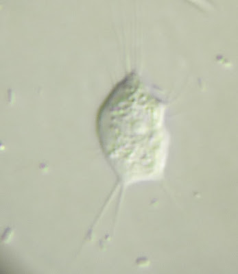
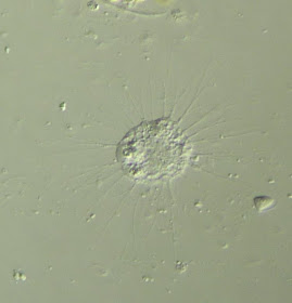
To begin, a fuzzy ciliate of sorts. Prominent contractile vacuole, and I think in the left image I think you can make out its macronucleus or two. Can't see the mouth so IDing it is a bit difficult.
[to shave off some page loading time, the rest is below the jump (if it works)]
This one's got a big mouth. Seems to be Lembadion sp., an oligohymenophoran ciliate. Guessing the yellowish particles are prey or food storage of some sort.
Epalxellid of sorts, perhaps of the genus Epalxella itself:
What appeared to be a developmentally-'unique' double stentor; or one in the process of division that remained attached. Actually, may well be division, didn't think they'd keep filtering away in the midst of cytokinesis:
Diplophrys, a stramenopile with a conspicuous lipid(?) droplet and very fine filopodia.
View of surface pellicle strips on some photosynthetic euglenid:
Some sort of glissomonad, I think. Looks hatchet/pike-like, would totally name it something silly like Hatchetomonas were it undescribed...
Some filose amoeba with seemingly eruptive lobopodia in addition to thin filopodia. Can nucleariids (amoeboid sister group to fungi) do this? Though the prominent nucleus at its centre points towards nucleariidness:
An amoeba with branching filopodia. Quite vacuolated. Fucked if I know what it is. Some sort of vampyrellid? (eg vaguely like this)
Nucleariid like the previously mentioned Rhabdiophrys? Or vampyrellid? Damn, identifying amoebae was way easier when I knew less of them...
Some small round thing that liked to wrap its [attached-to-substrate] flagellum around itself. Ummm, that sort of made sense, right? Don't think I'm allowed to ever write species descriptions for some reason...
"You really like amoebae" I was told the other week. Yes I do, and FYI, someone's chlorarachniophytes are amoebae too. All that said, I remain abysmal at identifying the buggers; this, my friends, is some filosey amoeba of sorts. Yup.
The following is from an anoxic rotting leaf sample (same as in the previous Trimastix post) – a very long and narrow cell, appears to be a euglenid based on how it moves the free flagellum (tip-heavy action); it's attached to the glass with the other flagellum. Guessing it's a Heteronema sp.; appears similar to H.acutissimum, but the latter is marine and for euglenids, the marine vs. freshwater distinction is generally considered sufficient to be a different species.
And now I'm off too look at more samples. Will take a while to go through my stockpile of protist pics already, but must still get more...


No comments:
Post a Comment
Markup Key:
- <b>bold</b> = bold
- <i>italic</i> = italic
- <a href="http://www.fieldofscience.com/">FoS</a> = FoS