Pages

Sunday Protist -- Oligotrich Ciliates: another morphological acid trip

Of course, no one noticed any delays in the posting of the Sunday Protist, because that never happened. Actually, I've been rather frazzled by this little fun activity that happens around this time of the year called 'finals', and thus had to desperately avoid any material I may find myself actually interested in, lest it hijacks my attention for too long. Also, I'll be mostly internetless starting tomorrow, and thus unable to blog. Coming back on 03 January. May or may not schedule a post, depending on time, but wish you all a very happy holiday and see you next year!

But before I take off, let's oogle at some ciliates. Don't have much time (last final tomorrow), so this post will be very superficial. Just sit back, relax and enjoy the Cornucopia of awesome strangeness that is ciliate morphology. Today's special features Oligotrichs, conspicuous by their tuft of cilia at one end.

Let's meet the prime representative, Strombidium:

Strombidium inclinatum; the weird frayed flat-looking things at the top are actually flagella linked together into polykinety, formerly known as a 'membranelle' for its membrane-like appearance in light microscopy. Those usually constitute the oral ciliature, which are involved in feeding. G is the girdle kinety, basically a single row of flagella wrapped around the cell. It has very few kinety (which, btw, consists of kinetIDs, or basal bodies. Try not getting them mixed up on a lab exam...) compared to something like Paramecium or Tetrahymena, which are covered in rows of kinety. (Modeo et al. 2003 JEM)

For a reminder of where ciliates are on the Big [sub]Tree of Life, they're alveolates here:

I was playing with tree software this weekend. And then, to cleanse myself of the shame, I downloaded a cellular pathway modeling program and played around with some networks. This is the scheme we go by at this blog, at the moment (see the list of organisms towards the bottom of the side bar -->) "Craptophyte" was shamelessly stolen from some real protistologists. Proper term is 'Hacrobia'.
[disclaimer]By the way, while alveolates+stramenopiles and amoebozoa+opisthokonts are pretty good together, the other branchings may or may not bear any resemblance to reality. Furthermore, the rooting of the tree is highly contested, but I'll go along with what TC-S says these days (what Tom says is quite subject to change though...), since it'll be harder for you to argue with me as you'd have to read his stuff yourselves. And most of you are too lazy to do that, so I'm safe. I am in no way liable for any damage or deaths that may result from the use of this tree. [/disclaimer]


Here's another sampler of Oligotrichs and their neighbouring Choreotrichs (which we should explore at a later date):

(Gao et al. 2009 Sys & Biodiv)
And how they relate to each other:

Oligotrich (and Choreotrich) phylogeny. We'll briefly look at Strombidium, Laboea and Pseudotontonia.(Gao et al. 2009 Sys & Biodiv)

I may have something oligotrich-like in this old sequence of images documenting a random ciliate exploding while I was taking an optical stack... the image quality is kinda crappy, so I could probably get away with calling it Strombidium or something. I don't even remember where this sample came from, may well have been marine, hence the explosion in water.

Let's wander about the Oligotrich tree some more.
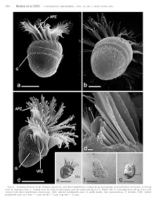
The first glimpse of Laboea made me double-check it wasn't actually some sort of tiny snail:

These SEMs are so nice I'm having trouble cropping them... (Agatha et al. 2004 JEM)

It also a rather unusual-looking (for a ciliate) cell division pattern:

Dividing Laboea sp. (Agatha et al. 2004 JEM)

If you've ever wondered how to tell a dividing ciliate from a conjugating couple, it's pretty easy: Ciliates divide transversely along the anterior-posterior axis, ie like <=<= if < style="font-style: italic;">Trichonympha is almost the polar opposite -- they divide laterally and have sex like this: <=<=. The Kama sutra of protists must definitely be written someday! See the bottom of this post for more ciliate sex.

Which is why Laboea looks a little weird, although here it must simply have a different longitudinal axis from what it first appears as. As much as I love morphogenesis/cellular development, and as much as tomorrow's final is Developmental Biol, ciliate morpho won't help much with chick gastrulation, sadly enough. Although it would be fun to draw parallels between various processes of development on morphogenesis to derive some fundamental principles, which I'm pretty sure (though only as much as a scientist can be sure about anything...) should repeat both on the unicellular and multicellular levels. After all, there's only so many ways to build a shape. So we'll have to pass on the cell division/morphogenesis stuff for now *sob*

Pseudotontonia. Ok, this thing is weird:

T - tentacles, TC - tail cilia. B shows a representation of the whole cell, which appears to have a very long 'tail' in addition to its tentacles. (Skovgaard & Legrand 2005 J Mar Biol Ass UK; free access)

To confirm that it really exists, there are pictures: (an SEM would be wonderful but this seems to be all we've got)

Pseudotontonia. Tail unfortunately not visible, although another image in the paper does show its beginnings. Fig F shows stained macronuclei. The thing is full of nuclear material. Ciliates have surreal genomes to match their similarly sophisticated cell structure. (Skovgaard & Legrand 2005 J Mar Biol Ass UK; free access)

Remember how Laboea has this weird division pattern, not very ciliate-like? To show how nothing makes sense except in light of evolution:

Proposed model of Oligotrich ciliary pattern evolution: The ancestor had multiple longitudinal rows of kinetids (basal bodies), which were subsequently reduced to two rows wrapping around the cell's main body. GK(red) - girdle kinety, VK(blue) - ventral kinety. c) Ventral kinety goes longitudinal. d) The anterior end of the girdle kinety went down to join its other end at the posterior (but not attach). e) The posterior end of the girdle kinety curved up along the ventral kinety. f) Posterior end of GK migrates upwards to form a circular girdle kinety at the anterior. g) Girdle kinety spirals down (eg. Laboea), ventral kinety disappears (Agatha 2004 Zool)

This helps make a bit of sense from the weird Laboea division -- the cells posterior (at least in terms of division) seems to be located elsewhere, messed up by the weird spiraling of the girdle kinety. The true posterior should be wherever the new cell starts to form.

Must...not...surrender...to morphology fetish... mmmm, ciliates! *drools* This is a really sexy paper on ciliary pattern evolution in Oligotrichs. Hmm, passing tomorrow's final, or really sexy paper. Shit. This is awful. I can't...no...can't look...stop me!

Actually, I'm gonna wrap it up there.

So, to summarise, (I need an excuse to put in this nice diagram):

Summary of Oligotrichs. Phylogeny based on morphological characters (indicated by numbers; explained in the paper). (Agatha 2004 Acta Protozool.; free access)

(I'm confused by the discrepencies between this tree, the Gao et al. 2009 tree at the beginning, and a few other Oligotrich/Spirotrich trees I've seen tonight, but with a final looming ahead in a few hours...I'll be a horrible blogger and pretend to ignore it for now! So here I present two sides of the argument. I hope the creotards creationists are happy.)

Ok, one last glimpse:

The arrow in C points to the organelle where the oral ciliature develops. Wonder if it's similar to the determinative region of oxytrichs... (eg. Grimes 1982) (Agatha 2004 Zool)

Wow. Ciliates definitely kick some dino ass. Take that, you-know-who-you-are! =P

Apologies to those who feel I may not have done justice to the works in this post; don't have time... also, warning: there will be no Sunday Protist next week. But I'll make up for it next year! =D

Happy holidays! Hiatus begins...now...

References
Agatha S (2004). A cladistic approach for the classification of oligotrichid ciliates (Ciliophora: Spirotricha) Acta Protozoologica , 43 (3), 201-217

AGATHA, S. (2004). Evolution of ciliary patterns in the Oligotrichida (Ciliophora, Spirotricha) and its taxonomic implications Zoology, 107 (2), 153-168 DOI: 10.1016/j.zool.2004.02.003

AGATHA, S., STRUDER-KYPKE, M., & BERAN, A. (2004). Morphologic and Genetic Variability in the Marine Planktonic Ciliate Laboea strobila Lohmann, 1908 (Ciliophora, Oligotrichia), with Notes on its Ontogenesis The Journal of Eukaryotic Microbiology, 51 (3), 267-281 DOI: 10.1111/j.1550-7408.2004.tb00567.x

Gao, S., Gong, J., Lynn, D., Lin, X., & Song, W. (2009). An updated phylogeny of oligotrich and choreotrich ciliates (Protozoa, Ciliophora, Spirotrichea) with representative taxa collected from Chinese coastal waters Systematics and Biodiversity, 7 (02) DOI: 10.1017/S1477200009002989

MODEO, L., PETRONI, G., ROSATI, G., & MONTAGNES, D. (2003). A Multidisciplinary Approach to Describe Protists: Redescriptions of Novistrombidium testaceum Anigstein 1914 and Strombidium inclinatum Montagnes, Taylor, and Lynn 1990 (Ciliophora, Oligotrichia) The Journal of Eukaryotic Microbiology, 50 (3), 175-189 DOI: 10.1111/j.1550-7408.2003.tb00114.x

Skovgaard, A., & Legrand, C. (2005). Observation of live specimens of Pseudotontonia cornuta (Ciliophora: Oligotrichida) reveals new distinctive characters Journal of the Marine Biological Association of the United Kingdom, 85 (4), 783-786 DOI: 10.1017/S0025315405011707

Mystery Micrograph #11

Shockingly enough (not really), the blogosphere's systematics god got the last one, which was Sporogena, a slime mould-esque ciliate! More on that sometime later, as I'm still rather swamped here (one more final). Let's have another one to guess. After Tuesday I won't be around to check comments until 04 Jan, so knock yourselves out. I intend to schedule random posts here and there in my absense, but may well not get around to it (other task 'debts' are more pressing...)

So here we go. Enjoy!

(to be referenced later)

Mystery Micrograph #10

Originally posted: 01.12.09 6:06PM
14.12.09 - bumped; solve it!

Johan got the last one -- it was Hoplonympha, a hypermastigote parabasalian; despite mischievously trying to look like Streblomastix (an oxymonad). If any or all of that sounded Greek/Latin to you, it's probably because it was it will be elucidated eventually, after I take care of the nematode foram parasite thing, as well as a delinquent Sunday Protist (which no one noticed, right? Good!)

Now let's look at something that appears more familiar:

Sadly, no scalebars (yay 1980s), so 18 - 200x; 19 - 230x; 20 - 200x; 21 - 170x; 22 - 1200x; 23 - 1800x (to be referenced later)

Also, to be fair, I did ask Opisthokont to pick a random clade, to give me MM ideas, and then explicitly said I won't do it. In the interests of fairness, the clade was...Breviata. So we know this is not a Breviate. Considering Breviates are this massive and famous group consisting of a tiny handful of species no one really cares about seeming to flip a finger at Tom's unikont idea (EDIT: Well, not entirely...), this should be the most useless hint to date.

Have fun! ^_^

HINT 13.12.09: The group of organisms this thing belongs to SHOULD NOT be doing this!
HINT 16.12.09: Perhaps this may help you a little.

(King 2004 Dev Cell; probably could add in a couple more lineages as 'multicellular-oid' but that's irrelevant...)

Sunday Protist - Phytomonas: plant trypanosomatids!

ResearchBlogging.orgWhile I was trying to come up with something quick to blog about, got a couple updates in Google Reader from J. Eukaryotic Microbiol, among them a paper on... trypanosomatids living in coconut tree phloem! Somehow, you don't typically think of plants being invaded by motile, flagellate things, but on a second thought: why not? The phloem is a vessel, and while perhaps there's no need to run away from macrophages or anything, there's no particular harm in retaining the ability to swim around. Especially if your other life happens in...insects!

Left: Phytomonas from coconut phloem. The arrow pointing to a transverse structure shows the sieve plate, which separate phloem tube cells. Note how the parasites congregate perpendicular to the plate. Kind of like salmon swimming upstream. The white round things in the middle image on the right are starch granules - food!(Keller & Miguens 2009 JEM; AOP)

For some idea of what these things are related to: (can I write a single post without showing or refering to a tree? Soon I'll get banished from organismal/cell biology...)

Phytomonas lives towards the bottom, amid monoxenous (single host) insect trypanosomatids. (Simpson et al. 2006 Trends Parasitol)

Trypanosomatids are worthy contenders for the Higher Parasite award (if ciliates are the higher eukaryotes, as we've established earlier, then why not have higher parasites as well?). In fact, they have a rather tight competition with the Apicomplexa, which are also a seriously effective bunch. Trypanosomatids and their brethren also have one of the most complicated mitochondrial genomes out there (if not THE most complicated), as alluded to towards the bottom of the Diplonemid post. They also have this nasty habit of constantly changing their surface proteins, thus outsmarting the host's immune response.

They can also be considered the reason why sub-Saharan Africa isn't Muslim, or particularly white for that matter: tryps are very good at completely decimating livestock, transport animals and clueless foreigners. Thus, the Islamic expansion was stopped upon reaching the Land of Tryps, as their camels and horses provided some much-needed fresh flesh for parasites, and running an empire without horses and camels is, well, difficult. Furthermore, Plasmodium and Trypanosoma did a nice job ganging up on the European invaders later on, both in person and by destroying their attempts at cattle farming. This story was told by a protistology instructor, demonstrating that protists can, in fact, dramatically impact human history. Ethnomicrobiology, the study of the interactions between humans and microbial life, would be a really cool thing to compile (and study)! Especially since almost every human culture on the planet has figured out a way to make their food rot in a way that it tastes nice, or sends you on a nice psychological trip. Usually the latter.

Again, to put things into morphological perspective, tryps are actually quite complex, despite what their simple wiggly appearance in light microscopy:

Overview of Tryp morphology, much more complex than the first impressions from microscopy. The review this is from discusses peculiar organelles called acidocalcisomes, which are apparently conserved throughout Eukarya and prokaryotes (eg. Agrobacterium), and may have been inherited from the bacterial proto-eukaryote. Seems to be involved in a whole bunch of biochemical ion pumping action. I personally prefer fun subcellular structures, like the cytoskeleton or the endomembrane system =P (Docampo et al. 2005 Nat Rev Microbiol)

The tryp flagellar pocket is a story in itself, even getting its very own review. Don't let the single flagellum fool you -- tryps are bikonts! The homologue of the inner flagellum in euglenids (which is really short already) simply got lost. Apparently screwing with flagellar structure really messes up the trypanosome cytokinesis, resulting in these wonderful convoluted clumps of parasite. But that's getting way off-topic...

Back to our trippy tryps. Tryps are predominantly insect parasites, but several lineages have taken a liking to vertebrates or plants on the side:

Monoxenous (single host) tryps spend their entire parasitic careers in insects. Phytomonas is heteroxenous, and alternates between insect and plant hosts, while Leishmania and Trypanosoma alternate between insects and vertebrates. Some insect tryps can also be found in plants, but I'm not too sure what exactly they mean by that. (Santos et al. 2007 Microbes & Infection)

Apparently some of those tryps don't particularly care whether they're hanging out in vertebrate blood vessels, insect haemolymph (or other organs) or plant phloem.
Turns out the haemolymph is quite low in oxygen levels, and I'd assume phloem sap would similarly not be anywhere near as rich as vertebrate blood. But on a second thought, much of the oxygen in vertebrate blood should be attached to haemoglobin, and thus not make much of a difference. What Tryps and co. are really after is glucose, which all three environments are rich in. Among other nutrients, of course, but here's a 2009 paper on Tryp energy metabolism for anyone who's into that sort of thing. *shudder*

So why infect plants? There's plenty of opportunity to accidentally learn to infect a new host if you spend a significant portion of your life (in vast numbers) resting as a spore outside your primary host. Thus, if your primary host happens to have a fetish for vertebrate blood, there's a high chance of frequent contact with that environment, and presumably it's similar enough to something the tryp is already adapted to. Thus, this jump to a new host isn't as shocking as it first looks. Similarly, if the insect host dines on plants, there's enough contact with the plant vascular system to eventually figure out a way to use it. After all, you might as well, especially since you don't have to be good enough to reproduce there or anything. There's a high likelihood of being slurped back up by the original host. So while really cool, it's not too shocking that such relationships evolve.

It would be interesting to trace host interactions of heteroxenous parasites (including fungi and oomycetes and all the rest); perhaps this host jumping is driven by a very close interaction between the two hosts. I know very little about the evolution of parasites, but it does seem really cool: how do the parasites (and other symbionts) manage to move between different hosts? Perhaps most often they simply coevolve with their host and diverge with them, but presumably cases of jumping between host lineages aren't all too rare?

Where this stuff could come in handy is that perhaps the infected plants may have evolved a nasty defensive response to Phytomonas. Phytomonas is a relative of Trypanosoma and Leishmania, which are not particularly welcomed by us, as they can be rather unkind (deadly). One wonders if anything can be learned from those plants and their defense strategies, and perhaps applied to human medicine. Somebody's probably on it already, I just don't follow biomedical literature.

Anyway, plant flagellate parasites = pretty awesome and unexpected. Upcoming biochem final = really UNawesome and quite expected. Anyone wanna write it for me?

Also, I owe posts and revised posts and other stuff. I'm on it, I swear! (and I really didn't mean to do a long Sunday Protist this time, but it always happens! Academic literature is like a freaking black hole/horribly addictive drug: sucks you right in, for hours! Or maybe I'm just insane... anyone else get sucked in for hours reading random papers on obscure topics? And actually enjoy it? Anyone?)

References:
Docampo, R., de Souza, W., Miranda, K., Rohloff, P., & Moreno, S. (2005). Acidocalcisomes — conserved from bacteria to man Nature Reviews Microbiology, 3 (3), 251-261 DOI: 10.1038/nrmicro1097

KELLER, D., & MIGUENS, F. (2009). In Vitro Cultivation and Morphological Characterization of Phloemic Trypanosomatids Isolated from Coconut Trees Journal of Eukaryotic Microbiology DOI: 10.1111/j.1550-7408.2009.00454.x

Santos, A., d'Avila-Levy, C., Elias, C., Vermelho, A., & Branquinha, M. (2007). Phytomonas serpens: immunological similarities with the human trypanosomatid pathogens Microbes and Infection, 9 (8), 915-921 DOI: 10.1016/j.micinf.2007.03.018

SIMPSON, A., STEVENS, J., & LUKES, J. (2006). The evolution and diversity of kinetoplastid flagellates Trends in Parasitology, 22 (4), 168-174 DOI: 10.1016/j.pt.2006.02.006

MM09 answer: Hoplonympha - loaded with bacteria

ResearchBlogging.orgI'm really behind on the answers. I'll do the easier one first, so MM08 will be next.

Remember this myserious organism from a while ago? Johan got it: it's Hoplonympha, a parabasalian gut endosymbiont! (Opisthokont was also on the right track)

Hoplonympha. top: SEM of whole organism (F indicates flagella), the long strips are actually ectosymbiotic bacteria, as evident in the TEM cross section on the bottom. CM - cytoplasmic [inner] membrane, OM - outer membrane, SL - S-layer. Note that unlike in Streblomastix (an oxymonad), the host cell is substantially more convoluted. (Image from Ohkuma 2008 Tr Microbiol(free access), originally from Noda et al. 2005 Env Microbiol)

A Streblomastix wannabe. Although in a completely different clade. Not too surprising, considering the similarity of their habitats, that such a strong evolutionary convergence may occur. Note that unlike Streblo, this organism also seems to contain bacterial endosymbionts inside it. It's quite a jungle of symbiotic relationships there in the termite and cockroach guts!

What are those bacteria doing? For one thing, Parabaslians are anaerobes, containing highly derived relict mitochondria called hydrogenosomes -- which, as their name suggests, generate hydrogen gas. Bare hydrogen is a relatively rare commodity in nature, so there's plenty of bacteria that crave it for their own metabolic exercises. Many of the symbiotic bacteria are methanogens, and use the hydrogen gas in their methane production pathways.

The exact functions of some other bacteria in this bizzare and complex ecosystem aren't well understood (Ohkuma 2008 Tr Microbiol). For many obligate anaerobes, however, the gut of various animals became a rare haven from the oxygen pollution their ancestors have wrecked the environment with a couple billion years ago. In termites and wood-eating roaches you have the extra advantage of free poorly digested (by the host) carbon sources entering in the form of wood cellulose. It's a nice deal: you nibble on yummy cellulose and the host is happy with your excrement. Of course, as with any nice deal, a hungry horde of other creatures congregates around the fun. So we end up with something like this:

A sample of the complex interactions between the gut protists, bacteria and the host. For more info, read the source Ohkuma 2008 Tr Microbiol, a freely accessible pdf of which was found by Johan.

And it's likely only the beginning of the story. And yes, the cellulose digestion is predominantly done by the protists, not the bacteria. Apparently, removal of bacteria by antibiotics did not stop the cellulose digestion, whereas a removal of the gut protists wrecks it.

Since it's meaningless to look at organisms without at least considering their place in The Tree, Hoplonympha seem to form a sister clade to Eucomonympha, which together group cozily with the Trichonymphidae, with some peculiar Staurojoeninidae getting in the way:
You may recognise Trichonympha, Eucomonympha, and Cochlosoma in the Trichomonads. Trichonympha are NOT Trichomonads, but are Hypermastigotes. Just sayin'. Trichomonads tend to be a little less 'hyper' with their karyomastigont (nucleus + flagellar apparatus) multiplication. Turns out we're steadily building up quite a collection of Parabasalians here...we have these people to partly blame: (Carpenter, Chow & Keeling 2009 JEM)

There's a really cool Parabasalian with ectosymbiotic bacteria that act much like flagella, propelling the organism by beating in a synchronised fashion. This partly where Margulis gets her "spirochaete = flagellum" fantasies from, where spirochaetes mafically became attached to the proto-eukaryote and somehow became its flagellum. Which is obviously eukaryotic, and devoid of DNA, and not even barely spirochaete-like, but never mind. Or, as TC-S would say: the eukaryotic flagellum differs from a spirochaete "in every visible respect possible" for a subcellular structure. =D We'll look at this cool organism at some other time, so I'll leave you in suspense for now.

Before I finally shut up, there's a slightly annoying gap in our exploration of Hypermastigotes: While we've by now glanced at Trichonympha, Eucomonympha and Hoplonympha, what about this mysterious Staurojoenina thing between them? Guess what, it also has ectosymbiotic bacteria!

Staurojoenina. The things on its ass in the SEM are spirochaetes, while the rest of it is covered in rod-shaped bacteria, with some flagellar tufts towards the anterior. Also littered with endosymbionts. Kind of cute, but Eucomonympha and Trichonympha are fuzzier. (Stingl et al. 2004 Microbiol)

Now to write up MM08 (a really cool one), and you guys still need to figure out MM10!

References:

CARPENTER, K., CHOW, L., & KEELING, P. (2009). Morphology, Phylogeny, and Diversity of
(Parabasalia: Hypermastigida) of the Wood-Feeding Cockroach
Journal of Eukaryotic Microbiology, 56 (4), 305-313 DOI: 10.1111/j.1550-7408.2009.00406.x


Noda, S., Inoue, T., Hongoh, Y., Kawai, M., Nalepa, C., Vongkaluang, C., Kudo, T., & Ohkuma, M. (2006). Identification and characterization of ectosymbionts of distinct lineages in Bacteroidales attached to flagellated protists in the gut of termites and a wood-feeding cockroach Environmental Microbiology, 8 (1), 11-20 DOI: 10.1111/j.1462-2920.2005.00860.x


OHKUMA, M. (2008). Symbioses of flagellates and prokaryotes in the gut of lower termites Trends in Microbiology, 16 (7), 345-352 DOI: 10.1016/j.tim.2008.04.004

Stingl, U. (2004). Symbionts of the gut flagellate Staurojoenina sp. from Neotermes cubanus represent a novel, termite-associated lineage of Bacteroidales: description of 'Candidatus Vestibaculum illigatum' Microbiology, 150 (7), 2229-2235 DOI: 10.1099/mic.0.27135-0

Divine masterpiece of a diagram...

In my quest to conquer as many TC-S papers as humanly possible, I came across this masterpiece of visual presentation*:

Love the "missing link" in the most prominent location. This is gonna be good...! (none other than TC-S, 2006 Biol Direct; open access)

83 page paper. 4 exams ahead. Tom's paper, wild hypotheses, bold assertions, *drools* cellular evolution. Exams, marks, future career. Hmmm. Damn you, Tom, I think you win this time...I will only read a few pages. The rest will be read over the holidays. I shall set my limit to...6 pages max until after the biochem final. And practise self-control. I CAN abstain from alcoholism drugs sex TC-S papers, I can! OMG, I has TC-S addiction... o_O

People in that field call me crazy. I can't quite figure out why...

Speaking of which, might anyone happen to have TC-S 2004 The membranome and membrane heredity in development and evolution
in Organelles, genomes, and eukaryote phylogeny: an evolutionary synthesis in the age of genomics Eds.: Hirt & Horner?
or his other chapter "Chromalveolate diversity and cell megaevolution: interplay of membranes, genomes and cytoskeleton"? We actually don't have that book, and there are no accessible PDFs out there, apparently. Alternatively, I could order it through inter-library loan, but I think I've harassed them enough for this year...


*His diagrams are 'special' enough to be featured on journal covers...

On a more personal note, I will try to abstain from blogging excessively over the next few weeks. Finals, etc, you see. 'tis the season to be cramming, lalala lala la la la la. Which is why I'm sitting here reading about eukaryotic megacell megaevolution. So if I'm falling behind on my posting obligations, that's why. In reality, I'm gonna be procrastinating my ass off, but we don't talk about that here. Perhaps I should write 1001 Things I'd Rather Do Instead of Cramming for Biochem Final. Would be such a bestseller. I mean, the possibilities are truly ENDLESS!

After finals, I'll have very sparse internet access until 04 Jan. Blogging will resume normally then.

That said, watch my post rate increase two-fold all of the sudden. And marks drop accordingly...

Speaking of marks, there's a guy in our lab who spent 5 years studying biology, and proudly brags about NEVER having stepped foot in a library all those years. His marks are really good.

Sunday Protist - Paramyxids: Nested parasites and introverted multicellularity

ResearchBlogging.orgA while ago we peered into the lidded jar-like spores of Haplosporidia, memorable by their peculiar habbit of building up pressure and popping open the lid upon germination, much like a jack-in-the-box. But nastier, if you're an oyster. While digging around in obscure haplosporidian literature, we came across their lesser known close relatives, the Paramyxids, characterised by their pechant for sporulating 'inward' several times, in a strange genre of parasitism reminiscent of matryoshka dolls*. Not only do they undergo serial endogenous divisions, Paramyxids exhibit differentiation among those cell types as well. Instead of being multicellular by growing outward like normal organisms, Paramyxids have evolved some strange sort of 'introverted' multicellularity.

Judging from these hints of a rather complex life cycle, one can probably predict Paramyxids to be parasitic, like their Haplosporidian relatives. Parasites tend to love convoluted life cycle gymnastics, perhaps because it's a wonderful way to trick biologists into considering the various stages to be different species, or even higher taxa. Speaking of which, quite a bit of time was spend trying to sort out their damn phylogenetic position - their identity as fellow Cercozoan sisters of Haplosporidia was not unanimously agreed upon. Since that discussion involves lots of technical phylogeny- and taxonomy-related ramblings, I've shoved that for later, at the very bottom of the post. Feel free to ignore, although I do think it is interesting.

Introduction to the Spore-in-a-spore-in-a-spore-in-a-spore-...
There are four genera: Paramyxa, Marteilia, Paramarteilia and Marteilioides, although Feist et al. 2009 propose supressing the latter to Marteilia. So let's pretend it doesn't exist. The life cycle starts off with an amoeboid primary 'stem cell', which crawls around between host cells and later gives rise to a secondary cell inside it. This cell then undergoes equal mitosis, giving rise to a second secondary cell. This part is common between all Paramyxids. The secondary cell (sporont) then divides endogenously to form one or several spores, depending on the species. Those spores divide endogenously again several times, up to the sixth generation (so three times) in Paramyxoides nephtys. To start off, the 'simplest' is Paramarteilia, with the spores dividing once endogenously:
TEM of Paramarteilia canceri, a crab parasite. The organism on the right is in an earlier stage of development. The labeling conventions in the vast field of Paramyxidology are as such: C and N indicate cell/cytoplasm and nuclei, respectively, with the number indicating the generation of endogenous division. So N1 is the nucleus of the primary cell (C1), and the cell immediately internal is indicated as C2, etc. This gets annoyingly confusing when we get to Marteilia, Paramyxa and Paramyxoides. This organism is quite simple, relatively, with two secondary cells (C2) each holding two spores (C3), each of which, in turn, has a single internal cell (C4). (Feist et al. 2009 Folia Parasitol)

Paramyxids are topologically taxing, as the 'simple' Paramartelia already begins to suggest. It gets distinctly worse, as nicely summarised in these two diagrams:

Paramarteilia (1), Marteilia (2), Paramyxa (3) share a few common early stages, consisting of an amoeboid stem cell crawling between the host cells. Upon the first division, this stem cell produces a couple secondary cells inside it, which later give rise to a number of spores, which themselves have a number of cells within. The latter two traits are species-dependent. The figure on the left shows the development in Paramyxa paradoxa, to be read clockwise. The intelligent designer was definitely high out of his mind when he made these.(Desportes 1984 Origins of Life)

Paramyxoides sporulation: a cytological acid trip
Let's take Paramyxa (and Paramyxoides, which for our purposes can be considered more or less the same thing) for a bit of a cytological acid trip. Paramyxa crawls about as a primary cell, divides endogenously once to make one secondary cell, which then divides 'normally' to form a second secondary cell, each of which then divides endogenously to form four spore cells (tertiary), which then divids inward another three times to form a nested spore thing (fig 3 below, A and B). Meanwhile, towards maturity, each of the spores forms an external sac, because it doesn't have enough membranes yet.

To further enhance its elaborate membrane collection, the fourth and fifth cells (so second and third cells within the spore), form haplosporosomes. (Annoyingly, I can't quite figure out what the haplosporosomes do. Perhaps nobody really knows, judging from their consistent description as 'electron-dense vesicles', even in recent literature.) Then, the primary and secondary cells disintegrate, releasing the spores. By the way, we're inside the host cytoplasm at this point. Then, the extrasporal sac gets smaller, and striated projections are formed which bind the four spores together. Said striated projections cement together the four spores into a tetraspore complex (fig 3 below, D). What happens after? We don't talk about that. Actually, I don't think anyone knows...

I got really confused so I made this diagram:
EM of a Paramyxoides (parasite of a polychaete) spore from Larsson& Koie (2005), with an attempt at representing the whole organism. Note the six generations of endogenous cell budding involved in this process. Cells 4 and 5 exhibit the haplosporosomes characteristic of Ascetosporeans. The primary cell usually degrades early on in development, while the sporont disintegrates later to release the spores. The spores secrete an extracellular membrane, which later shrivels up and forms mysterious 'striated projections', whose function appears to be cementing the spores together. (Since I have no EM experience, trust my membrane tracing attempts at your own risk. It involved more imagination than anything else, really...)

Cellular invasion -- somebody needs to do some work on this so I can expand this section
So then I was excited to read about the Paramyxid cell invasion mechanism, and later compare it to that of microsporidia and apicomplexa. Alas, just as for the sister Haplosporidia, the literature lies silent about the exact journey of the paramyxid into the host cytoplasm. It's mentioned that the parasite develops in direct contact with the host cytoplasm (Larsson& Koie 2005), probably implying that there's no parasitophorous vacuole or any such thing. Thankfully. I have no idea how nature would put up with one more membrane...

Paramyxid life cycles: Question marks and dotted lines
To confuse ourselves further, I picked up a couple life cycle diagrams. Universal Law mandates that parasites must have life cycles from hell, so here's one of them:

Parasitic life cycles from hell, exhibit 1: Marteilioides chungmuensis (oyster parasite). Presumably, the dotted line means "And then it magically ends up inside the host cell" or something to that effect. It seems that the cell undergoes several developmental programs, and thus several different series of endogenous divisions, to make life interesting. Or maybe the same sequence but only executed to completion within the oocyte. Regardless, there is an 'extrasporogony' stage where the parasite endogenously replicates, after which it enters the gonads and magically forces its way into the oocyte, where sporulation happens. With question marks and dotted lines in-between. (Itoh et al. 2004 Int J Parasitol)

The oyster parasite Merteilia sydneyi (Kleeman et al. 2002) also has a life cycle from hell, also with an extrasporogony stage (incidently, it was first found there, and subsequently in Marteilioides). It seems that the gill epithelium may be a safe place for the parasite to proliferate. Once Marteilia reaches the gut epithelium, where it will eventually sporulate, it seems to undergo a few more rounds of non-sporogenic endogenous division, possibly to proliferate itself throughout the digestive system. In fact, this stage results in an exponential increase of parasitic cells, so efficient that nearly all the digestive tubules are infected. Once enough of the epithelial surface is covered, the parasites invade the host cells and form spores there. Which then...somehow...do something. Presumably, some membranes break somewhere in the process.

Marteilia sydneyi life cycle. See text. (Kleeman et al. 2002 Int J Parasitol)

The gut cell invasion and subsequent sporulation happens synchronously, en masse. This is also when the host finally launches an immune response, although a little too late. Parasites often employ the stealthy ninja tactics of multiplying rapidly somewhere quiet, where they go unnoticed, and then suddenly attacking in vast hordes and completely overwhelming the host. Kind of like the 14 year periodical cicadas overwhelm their predators, or how the malaria parasites lyse the host cells synchronously, causing the characteristic waves of fever.

The Kleeman et al. 2002 paper is the first study attempting to characterise the early developmental stages of a paramyxid oyster parasite. While sporulation has been glimpes at, we are only beginning to get an idea of this organism's fascinating life cycle. New techniques like in situ hybridisation are allowing researchers to finally trace the ellusive complex life cycles of many parasites, without having to rely on obscure morphological traits to identify the organism. A lot of the parasitic life cycles (as well as some free-living ones) turn out to be more complex than previously thought.

Summary and introverted multicellularity.
In case anyone is interested in the finer taxonomy of this group, here's the latest classification according to number of secondary cells, spores and number of cells within a spore.
The coolest thing from all this? The whole idea of multicellularity happening endogenously rather than usual 'extroverted' way. In both cases, we have cellular differentiation happening by asymmetrical mitosis. In paramyxids, this mitosis is abnormal, ie the daughter cell ends up inside the mother. There's another group of organisms that do this. You may actually be familiar with them: flowering plants. Pollen formation involves endogenous budding as well, as you have a vegetative cell, which forms the pollen tube, and a generative cell, which in involved in fertilisation.

I'm perplexed by the cell biology of engogenous mitosis, and perhaps it may be similar to whatever happens in pollen formation. However, we may have enough material for today. In fact the following part isn't necessary, as it's a bit of a discussion about the phylogenetic position of the paramyxids. If you care, feel free to stay. We will be discussing a whole paragraph from the Book of Tom, which may be interesting if that's your 'thing'. So here you go, another obscure group of parasites has been brought to light, and hopefully not mangled too much in the process. Apparently, multicellularity can also happen 'introvertedly' -- who knew?

Phylogenetic Home: Snuggling with Haplosporidia in the land of Cercozoa
[Warning: We're going to increasingly bury ourselves deeper and deeper into technical phylogenetic details, random hypothesising, and readings from the Book of Tom. Proceed with caution. Emergency exit that way ---> ]
Naturally, back in the days of crown eukaryotes and the protozoan-metazoan divide, Paramyxids were especially confusing taxonomically. They seemed multicellular-ish, thereby tempting researchers to place them with something 'higher', like metazoan-y things. However, the presence of similar inclusions to those which the Haplosporidia are known for -- haplosporosomes -- alluded to their relationship even back in the 70's. (eg. this French paper compares various Haplosporidia with Marteilia) Sprague (1979 Mar Fish Rev) published a taxonomic paper wherein they were lumped with Haplosporidians under the phylum Ascetospora, described as the following:
Phylum IV. ASCETOSPORA ph. n. (ascet- Gr. asketos, curiously wrought. Refers to the strange and complex spore structure, recently revealed with the electron microscope.) Spore multicellular (or unicellular?), with one or more sporoplasms. without polar capsules or filaments; parasitic.(Sprague 1979 Mar Fish Rev)
Remember, this was back in the day when Protista was a massive taxonomic MESS consisting of random stuff clumped together based on superficial morphological appearances. Ultrastructural studies messed this up dramatically, but true hell happened once sequencing became more available. A bit of a disaster struck when it was decided that sequencing should be as easy as 1. grab ribosomal DNA 2. sequence 3. align 4. ??? 5. Profit. Well, things did get aligned, and a lot of the rDNA data was quite consistent. Unfortunately, in some cases, it was consistently wrong.

Paramyxids seem to have suffered from the same taxonomic affliction as microsporidia -- namely, extreme [intracellular] parasitism tends to drive genomes to do wonky things. Of course, wonky genomes tend to provide habitat for some rather non-conformist sequences for homologues of conservative conserved genes. Ribosomal DNA is no exception. As we've seen before, highly diverged sequences tend to group with each other due to Long Branch Attraction. You can probably guess where this is headed: Paramyxids were yet another ancient eukaryote, branching right after Microsporidia (Berthe et al. 2000). You'd think people would be a little more perplexed by the bulk of 'ancient' eukaryotes being parasites...of eukaryotes. Yeah.

After having spent a few hours earlier digging through some rather unhappy-looking trees to find what paramyxids actually are, I'll just settle for TC-S and Chao (2002, 2003) , who place them in "Ascetosporea" (Sprague 1979), sister to Haplosporidians. According to TC-S and Chao (2002, see fig.6), both ["excessively long-branch"]Paramyxids and Haplosporidia contain a characteristic Cercozoan SSU V6 terminal loop deletion. That and the haplosporosomes should be enough for out purposes. According to Tom, they seem to group with Phytomyxea, a clade of plant pathogens containing that nasty sworn enemy of the Brassicales, Plasmodiophora brassicae. He goes on to suggest a homology between Plasmodiophorid 'cored vesicles' and haplosporosomes, thereby forming a potential synapomorphy for a group he calls Endomyxa, a new subphylum of Cercozoa. Furthermore, he suggests chemically targetting those haplosporosomes/cored vesicles to treat some of the commercial afflictions their organisms cause. See, real men do all that within a single paragraph of one paper. Oh yeah.

I'm trying to figure out what those 'cored vesicles' are, and so far I just found these unconvincing things in fig8 here (if you're still here, and don't have access, 1. Holy crap you're awesome for hanging in there for so long! 2. It's your garden variety random vesicles. Nothing much to look at...also, I'm just rambling right now. Seriously, not missing out on anything =P). Oh, there's some interesting-looking 'osmiophilic bodies' in Polymyxa (fig 3). To be honest, a lot of things have mystery vesicles, so I'm a little of skeptical of claims like "oh hey, these random tiny blobs are homologous to those random tiny blobs!". Meh, he's right about 80% of the time, so let's pretend they are homologous, sure. See if I care.

(Hmmm, I wonder if there's anything interesting about the seemingly U-shaped nucleus in Plasmodiophora and the U-shaped sporoplasm nucleus noted in Paramyxa. Or if it's just some artefact. Or coincidence. Probably that. But still, someone wanna go check it out?)


I think I may be running out of steam now...

*[completely off-topic] (from first paragraph) While writing this, I managed to go from Paramyxids to the Novgorod Codex in about 20min. Yay Wikipedia! Ancient Russian is so damn hard to read, but I still find it amazing to be able to decipher some words and phrases, almost an entire milenium later! Interestingly, Slavic languages seem to have evolved slower than their Germanic counterparts -- not only is the language diversity in that family much lower (and mutual intelligibility quite a bit higher), but 1000 year old Slavic languages are easier for modern speakers to decipher than 1000 year old English texts for Anglophones (cf. Beowulf). How many mouse clicks am I from the socioeconomics of Fiji, and then succulents of New Mexico? Might even click our way to Higg's Boson if we keep this up...[/completely off-topic]


References
Cavalier-Smith, T., & Chao, E. (2003). Phylogeny of Choanozoa, Apusozoa, and Other Protozoa and Early Eukaryote Megaevolution Journal of Molecular Evolution, 56 (5), 540-563 DOI: 10.1007/s00239-002-2424-z

Desportes, I. (1984). The Paramyxea Levine 1979: An original example of evolution towards multicellularity Origins of Life, 13 (3-4), 343-352 DOI: 10.1007/BF00927182

Feist SW, Hine PM, Bateman KS, Stentiford GD, & Longshaw M (2009). Paramarteilia canceri sp. n. (Cercozoa) in the European edible crab (Cancer pagurus) with a proposal for the revision of the order Paramyxida Chatton, 1911. Folia parasitologica, 56 (2), 73-85 PMID: 19606783

ITOH, N. (2004). Early developmental stages of a protozoan parasite, Marteilioides chungmuensis (Paramyxea), the causative agent of the ovary enlargement disease in the Pacific oyster, Crassostrea gigas International Journal for Parasitology, 34 (10), 1129-1135 DOI: 10.1016/j.ijpara.2004.06.001

Kleeman, S. (2002). Detection of the initial infective stages of the protozoan parasite Marteilia sydneyi in Saccostrea glomerata and their development through to sporogenesis International Journal for Parasitology, 32 (6), 767-784 DOI: 10.1016/S0020-7519(02)00025-5

LARSSON IR, & KØIE M (2005). Ultrastructual Study and Description of Paramyxoides nephtys gen. n.,
sp. n. a Parasite of Nephtyscaeca(Fabricius, 1780) (Polychaeta: Nephtyidae) Acta Protozoologica, 44 (2), 175-187


SPRAGUE V (1979). Classification of the Haplosporidia Marine Fisheries Review, 41 (1-2), 40-44